
Scientific illustration | 3D modelling
These images are from a poster explaining the discovery behind Peter Doherty's
and Rolf Zinkernagel's 1996 Nobel Prize in Physiology or Medicine.
The poster was published in 1997 by the Australian Academy of Science.
See the Acrobat PDF samples page for full-size front and back PDFs.
New illustrations from a 2005 Nobel Prize poster are here.
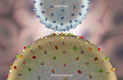
The killer T-cell is covered with a single type of receptor. The different coloured
spikes on the virus-infected cell represent different types of MHC antigens.
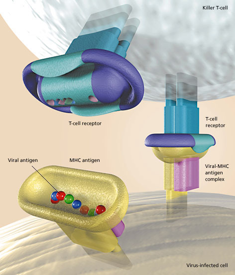
The surface of a killer T-cell has numerous receptors which are unique to a particular individual. A killer T-cell does not lock into an MHC antigen (healthy cell) without a viral antigen. When a viral antigen binds to an MHC antigen (virus-infected cell), the shape of the MHC antigen is altered and a matching receptor on a killer T-cell can recognise the resulting viral-MHC antigen complex. The T-cell receptor then locks into the viral-MHC antigen complex and the killer T-cell releases molecules that penetrate the infected cell and kill it. This shuts down the virus factory.
Technical notes
The basic geometry of the shapes was developed in several stages in Macromedia FreeHand. The resulting curves were imported into Alias Sketch, converted into three-dimensional objects, and cloned, modified and placed. Numerous test renderings and modifications were necessary for adjustments to the colours, lights, textures and transparency effects. The final ray-tracing of the larger image (500 x 425 mm at 225 dpi, file size 64 Mb) took over 12 hours (PowerMac 8500-132 MHz with 64 Mb of memory). Click here for a detail at full resolution.
All images and graphic elements in this site are protected by copyright
© 1996–2024 Paragraph®. All rights reserved.